Figure 6.
Formation of Anaphase Bridges in T1 35S:RNAi-GTBP1 Transgenic Cells.
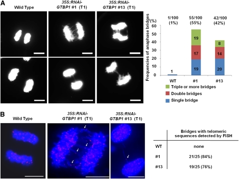
(A) Anaphase chromosomes from wild-type and T1 35S:RNAi-GTBP1 (lines #1 and #13) anther cells. Anaphase chromosome spreads were obtained from anthers of wild-type and 35S:RNAi-GTBP1 transgenic lines (#1 and #13), stained with DAPI, and observed by fluorescence microscopy. Abnormal anaphase bridges were detected in transgenic meiotic cells. Frequencies of single, double, and multiple anaphase bridges are indicated in the right panel. Bars = 10 μm.
(B) FISH analysis of anaphase chromosomes in wild-type and T1 35S:RNAi-GTBP1 (lines #1 and #13) meiotic cells using a (TTTAGGG)70 repeat telomeric fluorescent probe. Chromosomal DNA was denatured and incubated with Texas red-dUTP–incorporated (TTTAGGG)70 telomeric probe. The chromosomes were counterstained with DAPI and observed using fluorescence microscopy. Arrows indicate telomeric signals at the fusion points of anaphase bridges. Percentages of anaphase bridges with telomeric sequences detected by FISH are indicated in the right panel. Bars = 10 μm.

