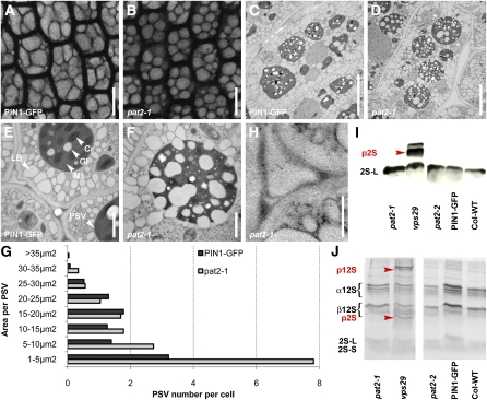
Figure 6.
pat2 Is Not Defective in the Trafficking of Storage Proteins to PSV.
(A) to (G) PSV autofluorescence (imaged immediately after peeling off the seed coat of the dry seeds) in root cells of control (A) and pat2-1 (B). Electron micrographs of control ([C] and [E]) and pat2-1 embryo root cells ([D] and [F]). Histogram evaluating the morphology of PSVs (n = 24 cells) (G).
(H) to (J) Immunogold labeling shows no accumulation of 2S albumin in the intercellular space of pat2-1 (H). Immunoblot (I) and SDS-PAGE (J) reveal no accumulation of 2S albumin and 12S globulin precursors in pat2 mutants, but they accumulate in the vps29, a mutant known to missort the reserve proteins.
Cr, crystalloid; Gl, globoid; Mt, matrix; p2S, 2S precursors; p12S, 12S precursors. Bars = 10 μm in (A) and (B), 5 μm in (C) and (D), 1 μm in (E) and (F), and 0.5 μm in (H).
[See online article for color version of this figure.]

