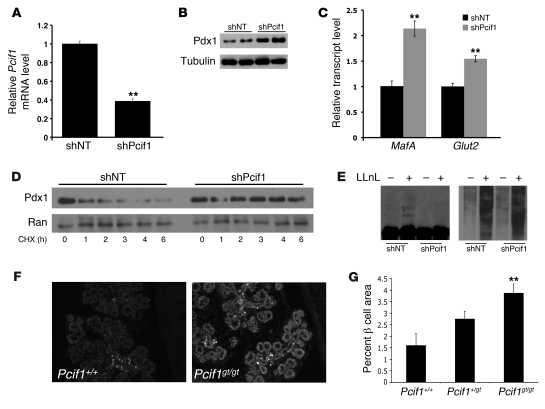
Figure 2. Increased Pdx1 accumulation and stability in Pcif1-deficient Min6 β cells and embryonic pancreas.
(A–C) Min6 cells nucleofected with control nontargeting shRNA (shNT) or shPcif1. (A) Pcif1 transcript measured by QPCR and normalized to Hprt. n = 3, **P < 0.01. (B) Western blot probed for Pdx1 and tubulin (loading control). (C) Transcript levels of Pdx1 transcriptional targets MafA and Glut2. n = 3, **P < 0.01. (D) Min6 cells expressing shNT or shPcif1, treated with the translation inhibitor cyclohexamide (CHX) and harvested at the time points indicated. Western blots are probed for Pdx1 and Ran (loading control). (E) Min6 cells overexpressing Cul3 and nontargeting shRNA or shPcif1 were treated with vehicle or 20 μM LLnL for 8 hours. Western blots are probed for Pdx1 (left) and ubiquitin (right). (F) Fluorescence staining of E16.5 embryos with dilute Pdx1 antibody. Original magnification ×10. (G) β Cell area of E18.5 embryos. n = 6, **P < 0.01 compared with Pcif1+/+.

