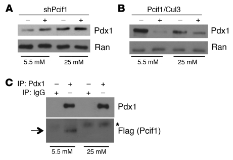
Figure 8. Posttranscriptional regulation of Pdx1 protein level by glucose.
(A) Western blot of lysates from Min6 cells maintained in 5.5 or 25 mM glucose, expressing nontargeting shRNA or shPCIF1. Blots are probed for Pdx1 and Ran (loading control). (B) Western blot of lysates from HEK293T cells maintained in 5.5 or 25 mM glucose, expressing Pdx1 in the presence or absence of overexpressed Pcif1 and Cul3. Blots are probed for Pdx1 and Ran. (C) HEK293T cells maintained in 5.5 or 25 mM glucose, transfected with plasmids expressing Pdx1 and Flag-Pcif1, and subjected to immunoprecipitation with IgG or Pdx1 antisera. Western blots are probed for Pdx1 or Flag (Pcif1). Arrow indicates migration of Flag-Pcif1; asterisk indicates heavy chain IgG signal.

