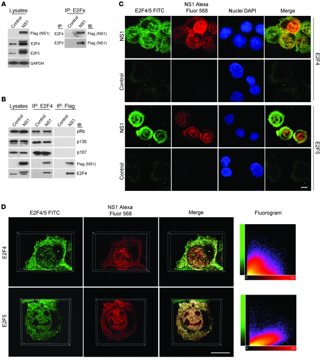
Figure 3. B19V NS1 protein enhances the nuclear import of E2F4 and E2F5 by formation of a heterocomplex.
NS1-transduced CD36+ EPCs were harvested at 24 hpt for subsequent experiments. (A) Whole cell lysates were subjected to immunoprecipitation by incubation with anti-E2F4 or anti-E2F5 antibody and then immunoblotting with anti-Flag (NS1) antibody. (B) Whole cell lysates were subjected to immunoprecipitation with anti-E2F4 or anti-Flag (NS1) antibody and subsequently analyzed by immunoblotting with the indicated antibodies. Whole cell lysates (5% input) without immunoprecipitation were also analyzed by immunoblotting as controls. (C) Cells were immunostained with antibody against E2F4, E2F5, or Flag (NS1), followed by secondary antibody conjugated with FITC (green) for individual E2Fs or with Alexa Fluor 568 (red) for Flag (NS1). After counterstaining of nuclei with DAPI (blue), cells were examined by confocal microscopy. (D) To address the relationship between E2F4/5 and NS1, z-series were collected throughout cells, and the 3D data were analyzed. Images were deconvolved, and the 3D renderings are shown. 2D fluorograms represent quantification of the colocalization of E2F4 or E2F5 with NS1: colocalization coefficients of 0.6 (E2F5 and NS1) and 0.3 (E2F4 and NS1) (1, perfect correlation; 0, no correlation; –1, perfect inverse correlation). Scale bars: 10 μm.

