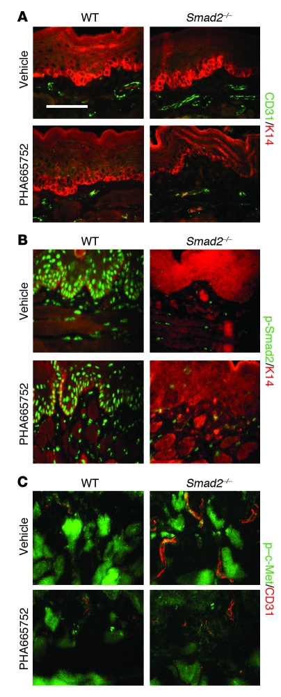
Figure 4. c-Met inhibition abrogates angiogenesis associated with epithelial Smad2 loss.
(A) IF staining of CD31 (green) shows increased angiogenesis in K5.Smad2–/– tongue mucosa, which was abrogated by a c-Met inhibitor PHA66572. K14 (red) was used for counterstain. (B) IF staining shows that PHA66572 did not affect Smad2 activation, as evidenced by staining of p-Smad2 (green). Note that p-Smad2 was specifically ablated in tongue epithelial cells but not in K5.Smad2–/– stroma. K14 (red) was used for counterstain. (C) Double IF staining of p–c-Met (green) and CD31 (red) shows that the vessels in K5.Smad2–/– tongue had increased p–c-Met staining (yellow or orange), which was abrogated by PHA66572 treatment. Bright green staining surrounding the vessels represents autofluorescence of the muscles in the tongue. Scale bar: 100 μm.

