
Figure 1.
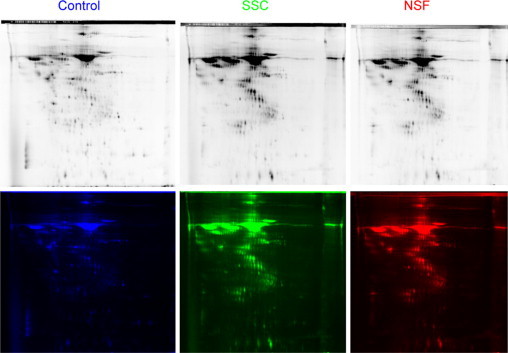
2D DIGE. The supernatants from three SSc, three NSF and three normal fibroblast cultures were pooled separately and analyzed. The secretome from normal fibroblasts was conjugated with Cy2 fluorochrome (blue), the secretome from SSc fibroblasts was conjugated with Cy3 (green), and the NSF secretome was conjugated with Cy5 (red). The top panels reflect the fluorescence intensity in each channel. The bottom panels represent the color assigned to each channel for visual differential analysis.