
Figure 5.
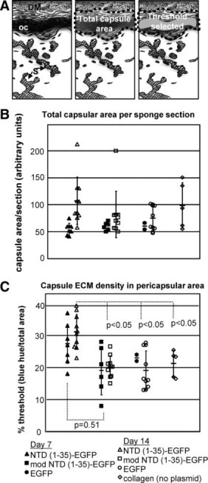
Collagenous ECM capsule density is increased in NTD (1-35)-EGFP sponges. A: Diagram of capsule quantification methodology. Images were captured along the entire pericapsular area of the sponge (left). DM indicates dermal muscle; oc, organized capsule; S, sponge material. The entire capsule region was selected (center) with the area containing organized collagen quantified based on the intensity of blue staining in Masson's trichrome–stained sections (right) using MetaMorph software and expressed as the percentage of the total capsular region that was designated as thresholded by these criteria. B: Total capsular area from sponges harvested at day 7 (filled symbols) and day 14 (open symbols). EGFP (circle), mod NTD (1-35)-EGFP (squares), NTD (1-35)-EGFP (triangles), and collagen only (diamonds). C: Collagen organization is expressed as the percentage of the total area defined by the threshold parameters. P values were calculated using the unpaired two-parameter Student's t-test.