
Figure 9.
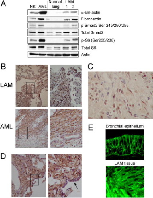
AML and LAM show evidence of EMT. A: Western blot analysis shows the expression of EMT markers (smooth muscle actin [α-sm-actin] and fibronectin), total and phospho (p)-Smad2, total and phospho-S6, and actin (loading control) in tissue lysates from human normal kidney (NK), AML, normal lung, and LAM samples. B: Immunohistochemical analyses of Snail in LAM and AML. Note nuclear staining in the abnormal cells. Right panels: high-power views of the boxed areas. C: High-magnification view of a LAM nodule showing heterogeneous expression of Snail, suggesting distinct populations of LAM cells. D: Immunohistochemical analysis of TGF-β in LAM tissue. Note the absence of TGF-β expression in normal epithelium (arrow in right panel [high-magnification]). E: Immunofluorescence analysis of E-cadherin in LAM compared with normal bronchial epithelium.