
Figure 2.
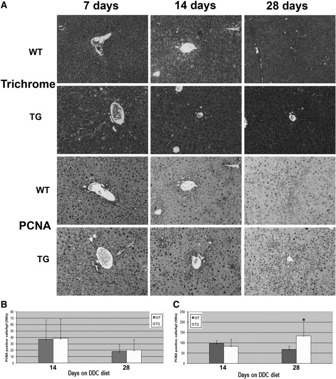
An increase in hepatocyte proliferation, but no change in fibrosis was observed in TG livers after short-term DDC exposure. A: Representative photomicrographs for trichrome (fibrosis, blue) and PCNA staining (proliferation, brown) of livers from WT and TG mice after 7, 14, and 28 days of feeding with the DDC diet. Original magnification, ×100. B: Quantification of PCNA-positive biliary epithelial cells at 14 and 28 days of DDC feeding shows no difference between WT and TG livers. C: Quantification of PCNA-positive hepatocytes at 14 and 28 days of DDC feeding shows a significant increase in TG livers at 28 days compared with WT livers. *P < 0.01.