
Figure 1.
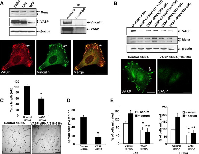
VASP is highly expressed in HSC, and VASP knockdown by specific siRNAs inhibits tubulogenesis, in vitro cell spreading, and migration. A, Upper left: Primary human HSC (HHSC), LX2, and MEF were subjected to Western blot analysis for Ena/VASP proteins. β-actin was used as a loading control. Upper right: Lysates of LX2 cells were subjected to IP using anti-VASP; coprecipitated vinculin was detected by Western blot analysis. Lower: LX2 cells were double-stained with anti-VASP (red) and anti-vinculin (green). Arrows mark colocalization of vinculin and VASP at a large and mature FA plaque. Scale bars = 20 μm. B, Upper: LX2 cells transfected with control siRNA or VASP siRNAs were lysed at 72 hours post transfection for Western blot analysis. The blot was reprobed with anti–β-actin as a protein loading control. Lower: VASP IF shows VASP was eliminated from FA and peripheral plasma membrane by VASP siRNA. Scale bars = 20 μm. C: Seventy-two hours post transfection, LX2 cells transfected with control siRNA or VASP siRNA were subjected to a tubulogenesis assay. Representative photomicrographs of tubes are shown on the bottom, and quantitative data are shown by a bar graph on the top (*P < 0.05, t-test; n = 3 independent experiments). VASP siRNA significantly inhibited vascular tubulogenesis of LX2 cells. Scale bars = 200 μm. D: Seventy-two hours post transfection, LX2 transfected with control siRNA or VASP siRNA were subjected to a cell spreading assay. VASP siRNA significantly inhibited cell spreading on culture dishes (*P < 0.05, t-test; n = 3 independent experiments). E: Cell migration was determined by Boyden chamber assays. VASP knockdown induced a similar inhibition of cell migration in both LX2 cell line and primary HHSC. *P < 0.05, VASP siRNA versus control siRNA in the absence of serum; **P < 0.05, VASP siRNA vs. control siRNA in the presence of serum.