
Figure 2.
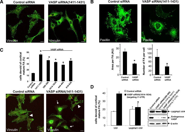
VASP siRNAs impair the development of mature FA. A: At 72 hours post transfection, confocal microscopy of vinculin IF (green) shows that FA signals were reduced by VASP siRNA(1411–1434). Scale bars = 20 μm. Both control and VASP siRNA were prelabeled with Cy3 (red). Scale bars = 20 μm. B, Upper: IF confocal microscopy of an alternative FA marker, paxillin (green), is reduced by VASP siRNA. Scale bars = 20 μm. Lower: High-power confocal images were subjected to FA analysis after background subtraction using Image Pro-Plus software. VASP siRNA significantly reduced the size of FA plaques and number of FA per cell in FA containing cells (*P < 0.05, t-test; n = 15 cells per group). C, Lower: Confocal images of vinculin IF reveal that VASP siRNA (1411–1434) increased the population of cells that are devoid of mature FA plaques (arrows). The arrowhead marks a single cell that still contains FA plaques. Scale bars = 20 μm. Upper: Five distinct VASP siRNAs exhibited a similar effect of increasing the population of cells that are devoid of FA plaques (*P < 0.05, analysis of variance; n = 3 independent experiments). D, Left: LX2 cells transfected with control or VASP siRNA (1814–1834) that targets against 3′-UTR were transduced with retroviruses encoding YFP or VASPWT-YFP. The effect of VASP siRNA (1814–1834) on FA was abolished by VASPWT-YFP fusion protein (*P < 0.05, analysis of variance; n = 3 independent experiments). Right: VASPWT-YFP fusion protein is resistant to targeting and degradation of VASP siRNA (1814–1834). Cells expressing VASPWT-YFP were transfected with various siRNAs, and Western blot analysis demonstrates that endogenous VASP but not VASPWT-YFP was degraded by VASP siRNA (1814–1834). As a control, both endogenous VASP and VASPWT-YFP were targeted by VASP siRNA (369–389), which targets against the coding region of VASP mRNA.