
Figure 5.
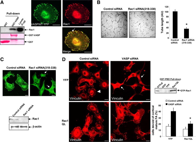
Constitutively active Rac1 reverses the FA phenotype of VASP siRNA. A, Left: GST-VASP interacts with Rac1 in vitro. GST or a GST-VASP fusion protein was incubated with LX2 cell lysates, and the bound Rac1 was detected by Western blot analysis. Ponceau S was used to assess purity of GST and GST-VASP fusion proteins. Right: Cells that express VASPWT-YFP were subjected to Rac1 IF (red). Arrow depicts colocalization of VASP with Rac1. Scale bars = 20 μm. B: At 72 hours post siRNA transfection, cells were harvested for tubulogenesis assays. Representative tubes formed on matrigel are shown on the left, and quantitative data are shown by a bar graph on the right. Scale bars = 200 μm. Rac1 siRNA (318–338) significantly inhibits vascular tubulogenesis of LX2 cells (*P < 0.05; t-test; n = 3 independent experiments). C: Confocal images of vinculin IF show that Rac1 siRNA(318–338) disrupts FA formation (upper, arrows). Scale bars = 20 μm. Lower: Western blot analysis show that Rac1 was efficiently knocked down by Rac1 siRNA (318–338) at 72 hours post siRNA transfection. D, Left: Cells transfected with control or VASP siRNA were subjected to retroviral transduction, respectively to express YFP or Rac1QL (constitutively active Rac1). Rac1QL reversed the FA phenotype of VASP siRNA. Arrowhead marks a single cell that still contains FA plaques. Arrows mark cells that are devoid of mature FA. Scale bars = 20 μm. Lower right: Quantitation of microscopy shows that Rac1QL significantly reduced the population of cells that are devoid of FA in VASP siRNA-transfected cells (*P < 0.05; analysis of variance; n = 3 independent experiments). Upper right: Highly increased Rac1 activity was detected in cells transduced with Rac1QL retroviruses by a Rac1 activity assay. Cell lysates pretreated with GTPγS or GDP were used as positive or negative control, respectively.