
Figure 6.
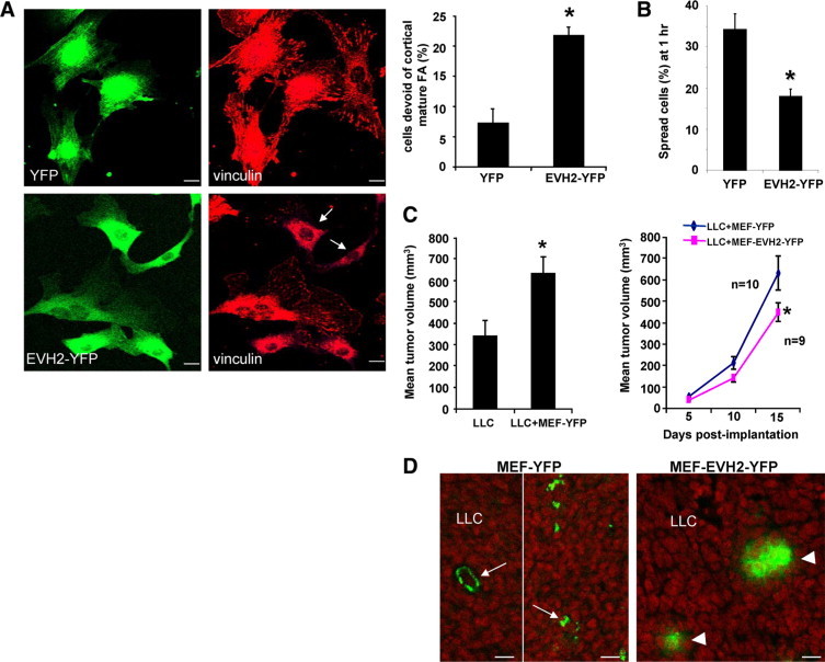
Perturbation of Ena/VASP function in MEF reduces the tumor growth conferred by tumor cell-MEF coimplantation. A: MEF expressing YFP or EVH2 mutant were subjected to vinculin IF. The EVH2 mutant induced impaired FA phenotype in MEF. Representative photomicrographs are shown in the left, and quantitative data are depicted by a bar graph in the right. Arrows mark cells that are devoid of large mature FA. Scale bars = 20 μm (*P < 0.05 t-test; n = 3 independent experiments). B: MEF expressing YFP or EVH2 mutant were subjected to a cell spreading assay. EVH2-YFP significantly inhibits cell spreading of MEF on cell culture dishes (*P < 0.05 t-test; n = 3 independent experiments). C, Left: Coimplantation of MEF with Lewis Lung carcinoma cells (LLC) subcutaneously into syngeneic mice promoted tumor growth compared with LLC alone (*P < 0.05 t-test; n = 9–10 tumors per group). Tumor nodules were measured on the 15th day post implantation. Right: 1 × 10⁶ LLC mixed with 1 × 10⁶ MEF that express either YFP or EVH2-YFP were implanted into syngeneic mice. Tumor nodules were measured and the tumor growth curves are displayed (*P < 0.05 t-test). D: Coimplanted YFP-tagged MEF were detected in tumor nodules on the 15th day post implantation by fluorescence confocal microscopy (green). MEF that express YFP incorporated into both vessels and interdigitation of tumor stroma (arrows, left). Conversely, MEF that express EVH2-YFP failed to distribute throughout the tumor stroma (arrowheads). Scale bars = 20 μm.