
Figure 5.
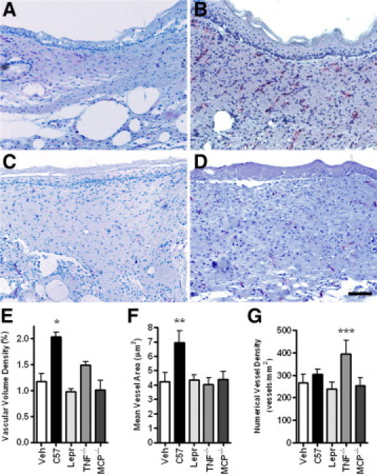
MCP-1−/− BMDCs do not induce vascularization of cutaneous wounds. Morphometric analysis of cutaneous wounds of Leprdb mice treated with vehicle (Veh), wild-type (C57), Leprdb (Lepr), TNF-α−/−, or MCP-1−/− BMDCs three days after the wound and harvested 14 days after wounding. Cutaneous wounds were serially sectioned, anti-CD31 immunolabeled, and examined at 140-μm intervals. A–D: Immunodetection of CD31 (red staining) in cutaneous wounds injected with vehicle (A), wild-type BMDCs (B), Leprdb BMDCs (C), or MCP-1−/− BMDCs (D). E: Vascular volume density expressed as percentage of wound volume. F: Mean vessel size (cross-sectional area) in wounded tissue. G: Numerical vessel density (number of blood vessels per area of injured skin). Note the correlation between the poor epidermal morphology and the lack of vascularization. *P < 0.05 relative to all groups except TNF-α−/−. **P < 0.05 relative to all groups. ***P < 0.05 relative to Leprdb. N = 4 to 8 per group. Error bars = SEM; Scale bar = 100 μm.