
Figure 7.
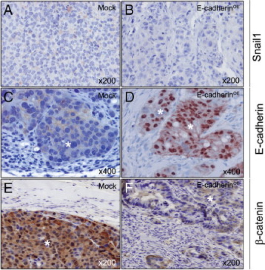
Snail1, E-cadherin, and β-catenin expression in peritoneal carcinomatotic foci of Mock and E-cadherinOE mice. Snail1 immunostaining was weak and cytosolic in Mock (A) and undetectable in E-cadherinOE (B) foci. E-cadherin immunostaining in peritoneal foci was weak and membranous in the Mock group (C) but very intense and mostly nuclear in the E-cadherinOE group (D). β-catenin immunostaining in carcinomatotic foci was very strong and mostly nuclear in the Mock group (E) but weak and cytosolic in the E-cadherinOE group (F). Hematoxylin costaining. White asterisks indicate tumor tissue.