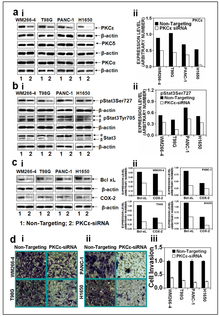
Figure 3. PKCε mediates phosphorylation of Stat3Ser727, Stat3-regulated genes expression and cell invasion in human cancer cells.
Melanoma (WM266-4), glioma (T98G), pancreatic (PANC-1), and lung (H1650) cancer cells were transfected with 15 µg of non-targeting siRNA plasmid (lane 1) or PKCε specific siRNA plasmid (lane 2) (Ambion, Austin, TX) for 48hr and whole cell lysates were prepared as described in Materials and Methods. The whole cell lysates (25 µg protein) were immunoblotted and indicated protein expression levels were detected with appropriate antibodies. β-actin was used as a control for gel loading variations. Protein quantification (normalized to β-actin) was done as described in Materials and Methods (right side). Expression levels of: a (i and ii), PKC isoforms (PKCε, PKCδ and PKCα), b (i and ii), pStat3Ser727, pStat3Tyr705, Stat3, and c (i and ii), Stat3 regulated genes (Bcl-xL, cdc25A and COX-2). d: Human cancer cell invasion. Cells were transfected with non-targeting siRNA plasmid or PKCε specific siRNA plasmid (Ambion, Austin, TX) and cell invasion was determined as described in Materials and Methods. (i): Photographs of invading cells. The migrant cells were stained with crystal violet and photographed the invading cells (40× magnification), (ii): Number of invading cells was estimated by colorimetric measurements at 560 nm according to assay instructions (Chemicon International, Temecula, CA). Each value in the graph is the mean ± S.E. from three separate wells.

