Fig. 4-A Fig. 4-B Fig. 4-C.
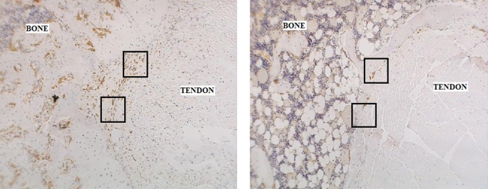
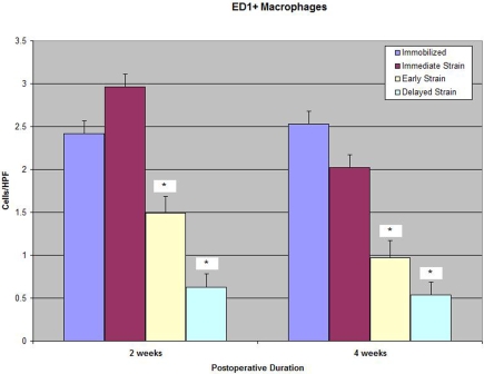
Fig. 4-A Immunohistochemical staining for ED1+ macrophages on an axial section of the tibial tunnel from an animal subjected to immediate loading and killed at four weeks (×40). Fig. 4-B Immunohistochemical staining for ED1+ macrophages on an axial section of the tibial tunnel from an animal treated with delayed loading starting on postoperative day 4 and killed at four weeks (×40). Note the substantially reduced quantity of ED1+ macrophages at the tendon-bone interface in the specimen from the delayed-loading group compared with that from the immediate-loading group. Fig. 4-C Significantly fewer catabolic ED1+ macrophages were observed at the tendon-bone interface of both delayed-loading groups compared with the number in the immediate-loading or immobilization group at two and four weeks postoperatively (p < 0.01). HPF = high-power field.


