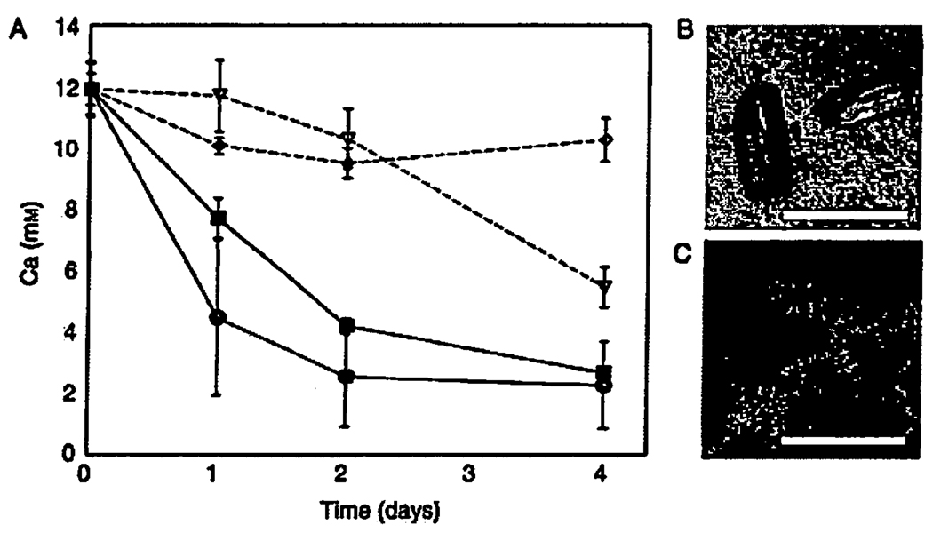
Fig. 2.
Removal of calcium from the photosynthetic growth medium stimulated by the photosynthetic activity of Rhodopseudomonas palustris. (A) Total calcium was measured by ICP-MS in triplicate samples. Black circles: an initial inoculum of 108 cells mL−1 at 7.8 µmol photon·m−2 · s−1. Black squares: an initial inoculum of 108 cells mL−1 at 3.8 µmol photon·m−2 · s−1. Empty triangles: an initial inoculum of 108 cells mL−1 wrapped in Al-foil. Empty diamonds: sterile control solution kept at the same distance from the light as the black circle samples, y-axis shows the concentration of calcium (mM) in the medium. Although the calcium concentration decreased in sterile controls, we did not detect any mineral precipitates either by light microscopy or XRD. (B) Transmitted-light micrograph of calcite crystals that grew in the presence of photosynthetica1ly active cells. (C) Top-view confocal micrograph of the epifluorescently stained cells (green/yellow) growing on crystals (red). The scale bar in B and C is 15 µm.

