Figure.
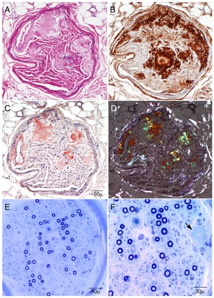
Superficial radial nerve biopsy (A-D, paraffin sections and E-F, epoxy sections). Serial paraffin cross sections show (A, hematoxylin and eosin stain) areas of eosinophilic amorphous deposits in the endoneurium and in the walls of endoneurial microvessels. The deposits react to lambda light chain preparations (B), stain salmon-pink on Congo red stain (C), and, when viewed under polarized light (D), show apple-green birefringence in the areas of amorphous material. The semithin epoxy sections (E and F) show a moderately reduced density of myelinated fibers. Unlike the clinical symptoms and findings (that are large fiber predominant), the biopsy shows relatively more severe reduction of small myelinated fibers in comparison to large myelinated fibers. The arrow shows a region of amyloid infiltration of an endoneurial microvessel. These findings are diagnostic of primary amyloidosis from a lambda light chain.

