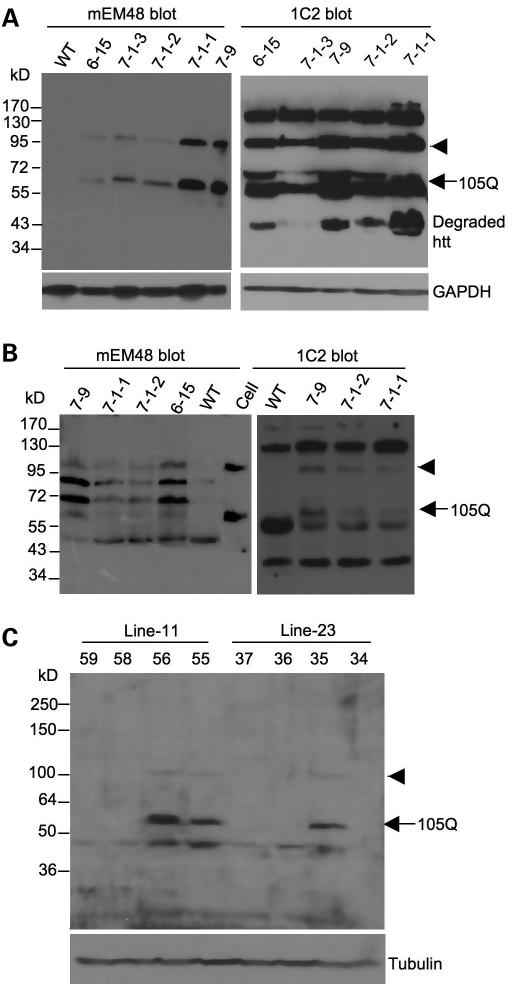
Figure 3.
Expression of mutant htt in transgenic HD pigs and mice. (A) Western blots of cultured fibroblast cells isolated from HD transgenic or wild-type (WT) pigs. The blots were probed with antibodies to htt (mEM48) and polyQ domain (1C2). Arrowhead indicates the uncleaved transgenic N208-105Q-F2A-ECFP protein. Arrow indicates the cleaved N208-105Q protein (105Q). (B) Western blotting of the brain cortical tissues from transgenic pigs (7-9, 7-1-2 and 7-1-3) and wild-type (WT) pig. Sample (Cell) from cultured fibroblast cells of a 7-1-2 pig was also included. Arrowhead indicates the uncleaved N208-105Q-F2A-ECFP. Arrow indicates the N208-105Q protein. The blots were probed with mEM48 for htt and 1C2 for the expanded polyQ tract. (C) Western blots of the brain cortical tissues from F1 transgenic HD mice (2 months old) of lines 11 and 23. Some mice (35, 55, 56) express the N208-105Q protein (arrow) and the uncleaved N208-105Q-F2A-ECFP protein (arrowhead), which are not seen in other littermates.

