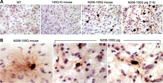
Figure 7.
Immunohistochemistry with an antibody to the activated form of caspase-3 of the mouse brain striatal tissues. (A) The brain striatum of wild-type (WT), HdhCAG140 knock-in (140Q KI), N208-105Q mice and N208-105Q transgenic pig (7–9) were immunostaininged with anti-caspase-3. Arrows indicate caspase-3-positive cells. (B) High power images showing caspase-3-positive neurons, which are distinct from negative cells and small glial nuclei. Scale bars: (A) 20 µm; (B) 10 µm.

