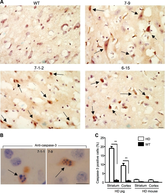
Figure 8.
Increased numbers of activated caspase-3-positive neurons in the brains of transgenic HD pig brains. (A) Increased numbers of caspase-3-positive cells (arrows) in the brain striatal tissues of transgenic HD pigs (7-9, 7-1-2 and 6-15) but not in wild-type pig (control). (B) Active caspase-3-positive neurons (arrows) in transgenic HD pig (7-1-1 and 7-9) brains show the DNA fragmentation feature of apoptosis. (C) Quantification of the relative number of caspase-3-positive cells in the transgenic HD pig and mouse brains. The images of HD mice are presented in Figures 4 and 7. The data were collected by examining the wild-type and HD (7-1-2, 7-9 and 6-15) pig brains and are presented as mean ± SE. **P<0.01 compared with control. Scale bars: 5 µm.

