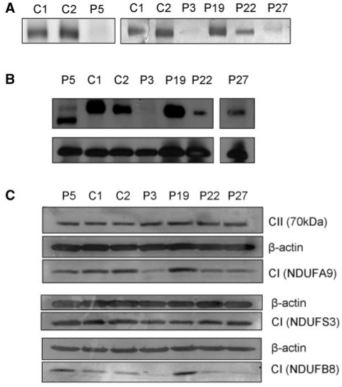
Figure 4.
Complex I in-gel activity and assembly in patient fibroblasts. (A) Complex I in-gel activity. Mitochondria-enriched protein fractions (75 μg) extracted from patient (P) and control (C) fibroblasts were separated on 5–13% 1D blue-native PAGE gels. Gels were histochemically stained for 2 h with 2 mM Tris–HCl (pH 7.4), 0.1 mg/ml NADH and 2.5 mg/ml nitrotetrazolium blue to assess complex I activity. (B) Complex I assembly. Mitochondria-enriched protein fractions (75 μg) extracted from patient and control fibroblasts were separated on 5–13% 1D blue-native PAGE gels. Proteins were transferred to a polyvinylidine fluoride membrane and probed with antibodies raised against complex I NDUFS3 and complex II 70 kDa subunits. (C) Complex I subunit expression. Total cellular protein lysates (10 μg) of patient and control fibroblasts were separated on 10% sodium dodecyl sulphate–PAGE gels then transferred to a polyvinylidine fluoride membrane. Western blotting was performed with antibodies raised against complex I subunits NDUFS9, NDUFS3 and NDUFB8, complex II 70 kDa subunit and β-actin.

