Figure 2.
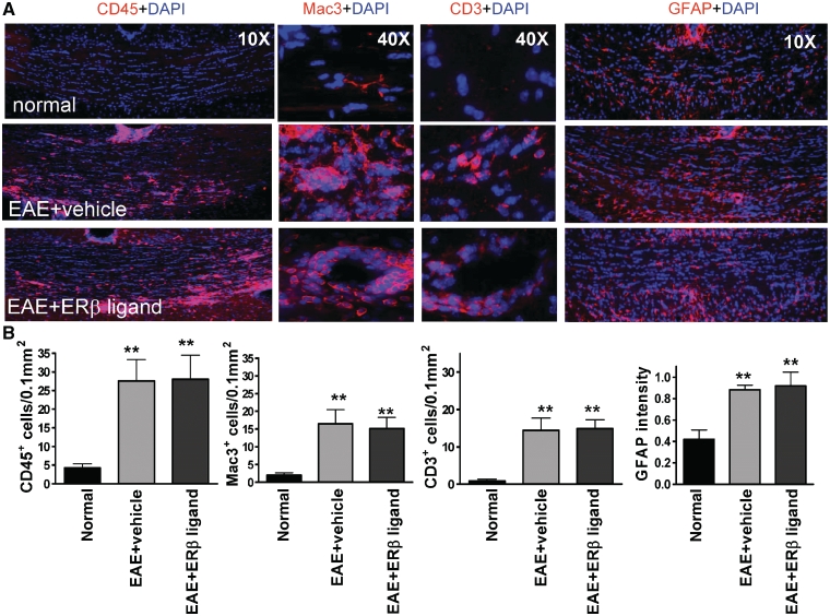
Treatment with oestrogen receptor β ligand did not reduce inflammation or reactive astrocytosis in the corpus callosum of mice with EAE. (A) Consecutive corpus callosum sections were also immunostained with antibodies against the common leukocyte antigen-CD45 (red, at ×10 magnification), the macrophage-Mac3 (red, at ×40 magnification), the T cell-CD3 (red, at ×40 magnification) or the astrocyte marker glial fibrillary acidic protein (red, at ×10 magnification). Shown are images from normal control, vehicle-treated EAE and oestrogen receptor β ligand-treated EAE corpus callosum at Day 36 after disease induction. Vehicle-treated EAE and oestrogen receptor β ligand-treated corpus callosum had large areas of CD45+, Mac3+ and CD3+ cells in the corpus callosum as compared to the normal control, as well as large areas of hypertrophic-reactive GFAP+ astrocytes. (B) Quantification of number of CD45+, Mac3+ and CD3+ cells and the relative fluorescence intensity of glial fibrillary acidic protein immunostaining demonstrated an increase in both vehicle-treated EAE and oestrogen receptor β ligand-treated EAE as compared to normal mice. Statistically significant compared to normal (**P < 0.001 ANOVAs; Bonferroni’s multiple comparison post test; n = 8–10 mice in each treatment group).

