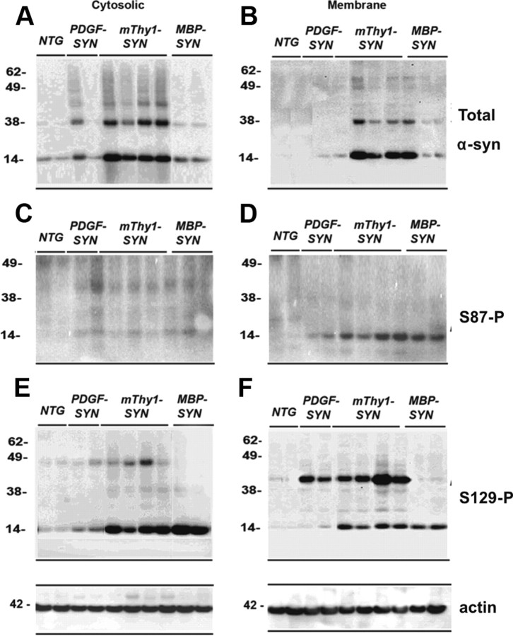
Figure 4.
Comparison of the levels of phosphorylated α-syn immunoreactivity by immunoblot in brains of α-syn models of LBD. Samples from the neocortex were divided into cytosolic and particulate fractions and analyzed by Western blot with antibodies against total and phosphorylated α-syn. The PDGF-α-syn express moderate levels of α-syn in the neocortex and hippocampus, the thy1-α-syn expresses high levels of α-syn in cortex and subcortex, the MBP-α-syn accumulates syn in oligos and is a model for aspects of MSA. A, B, Cytosolic and particulate fractions probed with an antibody against total α-syn. Native α-syn is identified at 14 kDa. In the TG mice there is an increase in native α-syn compared to non-TG. Compared to non-TG, in thy1-α-syn cases there was increased accumulation of α-syn aggregates above 28 kDa preferentially in the membrane fraction. C and D, S87-P α-syn was identified as a single band at 14 kDa. This band was exclusive to the membrane fraction and detected only in the TG mice cases. E, F, The native S129-P α-syn was identified as a single band at 14 kDa and oligomerized α-syn was identified as multiple bands ranging from 28 kDa to 98 kDa. The aggregated S129-P α-syn identified in the α-syn TG mice was more abundant in the membrane fraction. The native form was more abundant in the cytosolic fraction.

