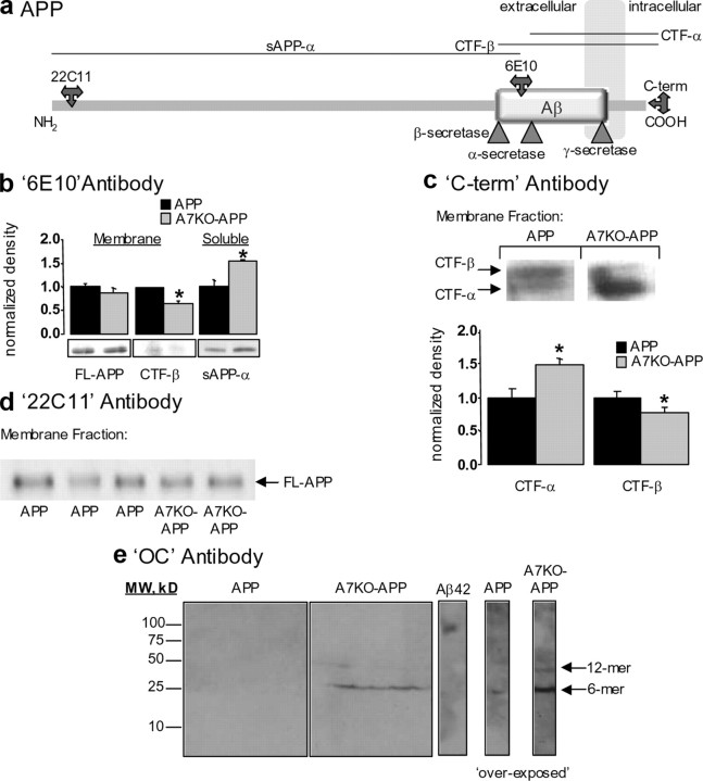
Figure 4.
Enhanced accumulation of soluble, fibrillar, oligomeric Aβ and altered APP processing in A7KO–APP hippocampus. a, Schematic depicting the APP within the cell membrane. Antibody epitopes and secretase cleavage sites are designated. b, Immunoblot with 6E10 antibody detects full-length APP (FL-APP) transgene and CTF-β in hippocampal membrane extracts from 5-month-old mice. Compared with APP mice, the FL-APP expression is unchanged (p > 0.05, Student's t test), but CTF-β is decreased in A7KO–APP samples (p < 0.05, Student's t test). The soluble product of α-secretase cleavage of the APP (sAPP-α) is enhanced in A7KO–APP hippocampus as detected with 6E10 immunoblot of soluble protein fraction (p < 0.05, Student's t test). Data expressed normalized to APP. c, Using an antibody directed against the C-terminal end of the APP indicates increased CTF-α and decreased CTF-β in A7KO–APP hippocampus. Left, Immunoblot; right, quantification of immunoblot results. *p < 0.05, Student's t test. Data expressed normalized to APP. d, Immunoblot with 22C11 antibody illustrates that amyloid precursor protein (holoprotein) expression levels in membrane fractions is unchanged between APP and A7KO–APP. e, Immunoblot with OC antibody demonstrates that A7KO–APP contain two fibrillar, oligomeric amyloid species,. The dodecamer species is absent from APP hippocampus as evidenced by an overexposure of the immunoblot. Positive control Aβ42 oligomer preparation is immunopositive for OC antibody with molecular weight (MW) of ∼120 kDa. Image shows APP and A7KO–APP samples with positive Aβ42 control cropped from the same immunoblot scan.

