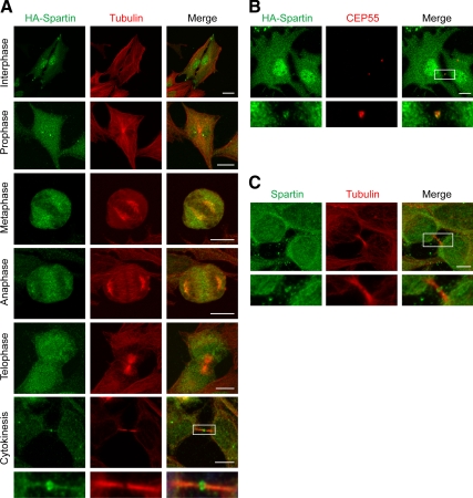
Figure 4.
Spartin localizes to midbodies during cytokinesis. (A) HA-spartin (green) accumulates at the midbody during late cytokinesis. Corresponding images show the localization of β-tubulin (red), and merged images are at the right. The boxed area is enlarged in the panels below. (B) HA-spartin (green) localizes to the midbody, as shown with costaining for CEP55 (red). The merged image is at the right. Boxed area is enlarged in the panels below. (C) HeLa cells were stained for endogenous spartin (green) and β-tubulin (red), with the merged image at the right. Boxed area is enlarged in the bottom panels. Bars, 10 μm.

