Figure 8.
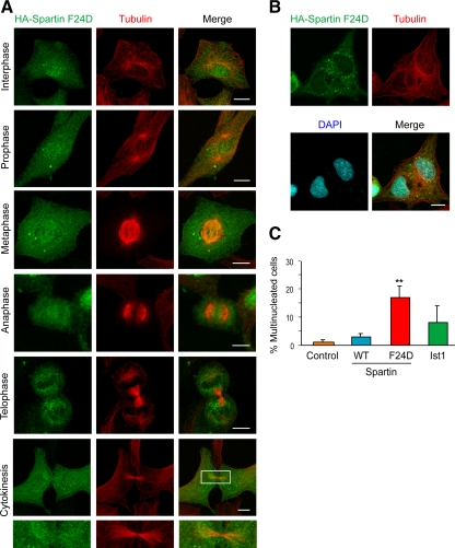
Spartin F24D does not localize to midbodies and causes dominant-negative impairment in cell division. (A) HA-spartin F24D (green) does not accumulate at the midbody during cytokinesis in HeLa cells. Corresponding images show the localization of β-tubulin (red), and merged images are at the right. The boxed area is enlarged in the panels below. (B) Multinucleated cells, with nuclei identified using DAPI staining (blue), were frequently seen upon HA-spartin F24D expression (green). β-Tubulin staining is shown in red. (C) Quantification of the percentage of multinucleated cells upon expression of empty vector, HA-spartin, HA-spartin F24D, and HA-Ist1 (means ± SD of at least three trials, with 100 cells/experiment). **p < 0.001 for wild type (WT) versus F24D. Bars, 10 μm.

