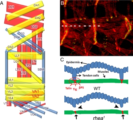
Figure 1.
(A) Schematic representation of the SMs in each abdominal hemi-segment A2–A7 of the developing embryo (lateral view with anterior left and dorsal up) by using the nomenclature of Bate (1993). Inner, middle, and outer muscle layers are shown in yellow, blue, and red, respectively (Bate, 1993). Dorsal oblique (DO), DA, dorsal transverse (Swan et al., 2004), LL, LO, LT (Cluzel et al., 2005), SBM, VL (Lundstrom et al., 2004), VA, VT, and VO. VA1 and VA2 are highlighted in red. (B) Muscle–muscle and muscle–tendon cell junctions in wild-type embryos visualized by staining developing muscle cells with actin (Tadokoro et al., 2003) and βPS integrin (green). (C) Diagrams showing a cross-sectional view along the broken line in B. The adhesion proteins (talin, βPS, and Tig, etc.) all concentrate at the end of SMs and are involved in forming stable muscle–muscle or muscle–tendon cell adhesions in wild-type embryos. In rhea1 mutant embryos, the muscle–tendon cell connections are broken (arrowheads), but the muscle–muscle connections remain (arrows).

