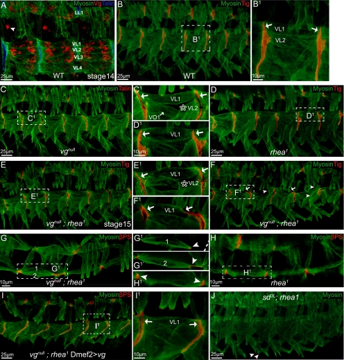
Figure 2.
SMs were detached in vgnull; rhea1 embryos but not in SD3L; rhea1 embryos. Embryos (stage 16 or a specified stage) are shown as lateral views, with dorsal up, and anterior to the left. Staining is color coded and indicated on each panel. B1–I1 are the close-ups of the framed area in B–I. (A) vg is expressed in muscle LL1 and VL1–4. The arrowhead points to a neuronal cell also expressing vg. Compared with wild-type embryos (B), vgnull (C), or rhea1 single mutation (D), or vgnull; rhea1 double mutant embryos in early stages (before stage 15; E), all produced a muscle pattern similar to wild-type embryos, except that a vgnull mutation caused muscle VL2 to be missing in ∼30% of segments (star in C1 and E1). Notice VL muscles (e.g., VL1) all formed tight adhesions between each other (arrows in B1–E1). (F–F1) By late stage 16 when muscles start to contract, muscle VL1 or other VL muscles detached from the attachment sites only in the vgnull; rhea1 double mutant embryos (arrows in F1). Arrows in F indicate detaching muscles and arrowheads indicate detached muscles. (G–H1) Overview of the vgnull; rhea1 double mutant embryos (stage 16; G–G1′) compared with rhea1 embryos (H–H1). Both VL1 and VL2 are retracting from their normal attachment sites (arrowhead in G1–H1). G1 and G1′ are two different confocal sections, and the broken line in G1 indicates the segment border. (I–I1) The muscle detachment phenotype of vgnull; rhea1 embryos can be rescued by expression of Vg via Dmef2-GAL4. Notice VL1 muscles built tight adhesions between each other (arrows in I1). (J) SD3L; rhea1 embryos did not have a muscle detachment phenotype. Some muscles do not develop well (VO4–6; arrowheads) in these embryos, but this mirrors the phenotype seen in SD3L single mutants.

