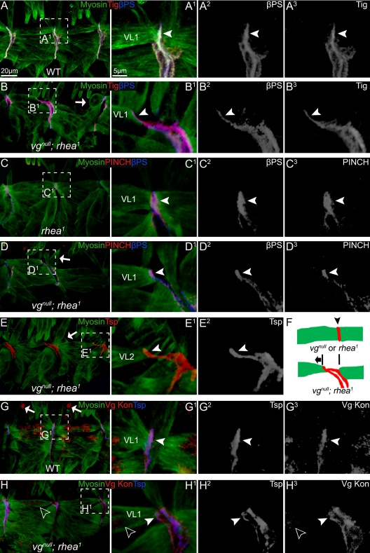
Figure 4.
The muscle detachment phenotype observed in vgnull; rhea1 embryos was not due to lack of localization of integrin or its known ligands, nor to an obvious muscle migration defect. (A) In wild-type embryos, βPS and Tig can be seen localized normally at the junctions between two VL muscles (arrowhead). (B) In vgnull; rhea1 double mutant embryos, the VL muscles were either detaching (arrowheads) or were already detached (arrows). However, βPS and Tig remain concentrated at muscle termini and followed the detaching muscles (arrowheads). (C) In rhea1 mutant embryos, the adhesion proteins PINCH and βPS formed tight junctions between VL muscles (arrowheads). (D) Similar to βPS and Tig, in detaching muscles in vgnull; rhea1 embryos, PINCH and βPS remain concentrated at muscle termini and followed the detaching muscles (arrowheads). Many muscles seemed to be detaching from the posterior border of each segment. (E) In the vgnull; rhea1 embryos, Tsp shows the same localization to the end of detaching muscles as PINCH, βPS, and Tig. (F) A diagram of the localization of adhesion proteins (red; arrowhead) in vgnull or rhea1 mutant embryos and the direction (anterior, arrow) in which VL muscles are moving after they detach. (G) In wild-type embryos, Kon, the major migration guidance protein for VL muscles, normally found at the end of muscle cells (arrowhead). (H) In vgnull; rhea1 embryos, some residual (maternally supplied) Vg protein can still be seen in VL1 muscle (empty arrowheads). These muscles still had a detachment phenotype, but Kon is localized properly (arrowhead). A1–H1 are the close-ups of the framed area in A–H. A2–H2 and A3–H3 show each confocal channel separately.

