Figure 7.
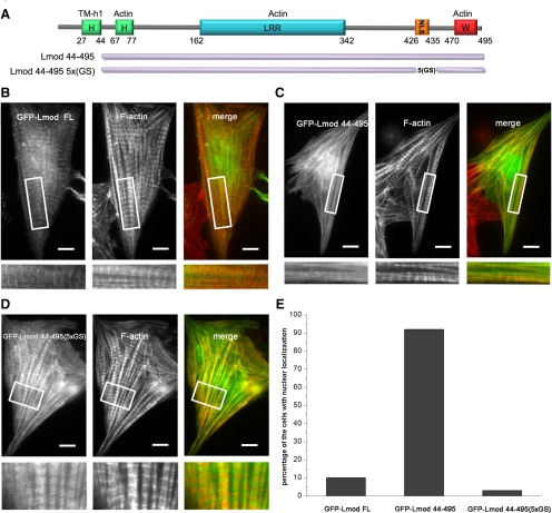
Subcellular localization of Lmod mutants lacking the tropomyosin-binding site and the basic patch. (A) Schematic representation of the GFP-Lmod constructs used in this study. Localization in cardiomyocytes of GFP-tagged LmodFL (B), Lmod44-495 (C), and Lmod44-495(5xGS) (D). Insets display selected regions of the cells at higher magnification, highlighting the localization pattern of each protein in myofibrils. LmodFL shows striated localization along myofibrils, with distinguishable accumulation near M-lines. Lmod44-495 shows diffuse localization along myofibrils, with a large fraction localizing to the nucleus (E). Lmod44(5xGS) displays a striated pattern along myofibrils but, in contrast to LmodFL, it does not accumulate near M-lines. The cells were also stained with rhodamine-phalloidin to visualize F-actin. Bars, 10 μm.

