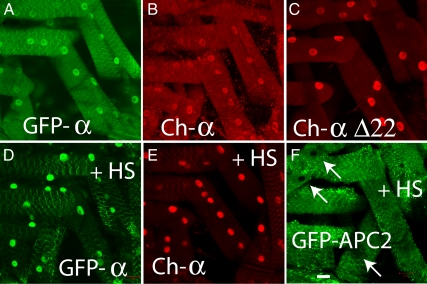
Figure 6.
Heat-shocked transgenic larvae demonstrate increased nuclear localization of AMPKα in vivo. Live animal images of 3rd instar Drosophila larvae expressing GFP-tagged (A and D) or mCherry-tagged wild-type AMPKα (B and E), mCherry-tagged truncated AMPKαΔC (C), or GFP-tagged APC2 (containing both NLS and NES sequences). Animals in D–F were subjected to heat shock at 37°C for 1 h, followed by a 15-min recovery at 25°C. All transgenic proteins are expressed using the Gal4-UAS system, driven by Ubiquitin-Gal4. Bar, 20 μm.

