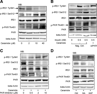
Figure 2.
PKR mediates the effects of ceramide on the phosphorylation of IRS1. HepG2 cells were exposed to different levels of ceramide for 12 h (A). Reverse transfection of suspended HepG2 cells was performed with scrambled siRNA (negative control) or siRNA of PKR for 24 h, and the transfected cells were cultured in regular media for another 12 h (B). Cells were then treated with ceramide (10 μM) for 12 h followed by insulin (0.5 nM) treatment for 15 min (B). Pretreated with 10 μM ceramide for 12 h, HepG2 cells were exposed to different levels of PKR inhibitor dissolved in DMSO (control; C) or 10 mM 2-AP dissolved in PBS:glacial acetic acid (200:1; GA, control; D) for another 12 h. After treatment, the cells were harvested, and Western blot analysis was performed to detect the level of β-actin and the total and phosphorylated levels of PKR and IRS1.

