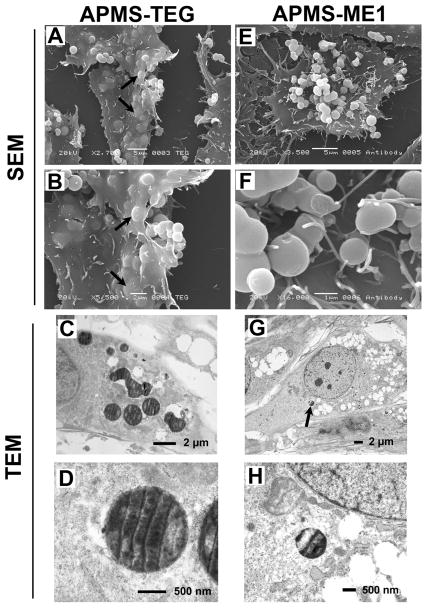
Figure 2.
Images of particles labeled with either TEG (APMS-TEG, A-D) or anti-mesothelin (APMS-ME1, E-H) interacting with cells 4 h after their addition to MM cells. SEM images showed that only particles carrying the TEG functional group were internalized by cells (arrows in A and B). APMS-TEG particles directly exposed to cytoplasm were observed in TEM (D). The arrow in image G indicates the lone APMS-ME1 particle found within an MM cell; it is enlarged in image H.

