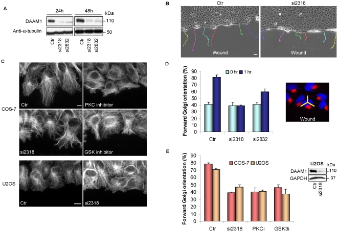
Figure 4. DAAM1 knockdown affects directed cell migration.
(A) Efficacy of two different siRNA against DAAM1 in COS-7 cells by Western analysis at 24 hours and 48 hours post-transfection. (B) COS-7 cells transfected with control (Ctr) or human DAAM1 siRNA (si2318) for 48 hours were scratched and observed using time-lapse imaging for 12 hours. Tracks of individual cells are shown as colored lines. Bar = 40 µm. (C) Wound-edge COS-7 cells subjected to GSK inhibitor (SB216763, 20 µM) and PKC inhibitor (RO-320432, 20 µM), or si2318 (40 pmol) were stained with anti-α-tubulin to visualize the microtubule network. Loss of DAAM1 was associated with random protrusions versus the more organized broad extensions in controls. Wound-edge U2OS cells subjected to siRNA treatment are shown in the bottom panel. Bar = 10 µm. (D) Forward Golgi orientation of COS-7 cells transfected with control (Ctr) or DAAM1 siRNA (si2318 or si2832, 40 pmol) at 0 and 1 hour post-wounding. Cells were scored for Golgi re-orientation using a 120° sector centered on the nucleus as shown on the right. (E) Right: Western analysis of DAAM1 knockdown in U2OS cells. Left: Golgi re-orientation in COS-7 and U2OS cells treated with various inhibitors 1 hour before wounding. PKC inhibitor (PKCi) and GSK inhibitors (GSKi) were used as in (c). In these analyses, cells were scored for Golgi re-orientation in three separate experiments. Error bars indicate standard deviation from the mean.

