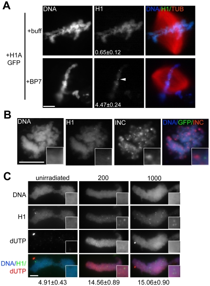
Figure 3. RanBP7 Reveals H1 Foci.
(A) Identically-scaled fluorescence images of fixed metaphase spindles from CSF reactions supplemented with 1 µM H1A-GFP and 4 µM RanBP7 or buffer control (+buff). In the presence of RanBP7, H1A-GFP is reduced on chromatin and concentrates on chromatin in small foci (arrowhead). The number of foci per nucleus (average ± standard error, n>40) is shown in the H1A-GFP column. (B) H1A-GFP foci do not colocalize with the centromere marker INCENP (INC). INCENP localization was performed using replicated chromosomes, on which H1A-GFP foci were less obvious but still detectable (insets). (C) Immunofluorescence images of UV-irradiated or unirradiated sperm nuclei assembled into chromatin in metaphase extracts supplemented with H1A-GFP, RanBP7, and biotin-dUTP. Insets are provided and the number of foci per nucleus is shown below the column for each condition. Scale bars, 10 µm.

