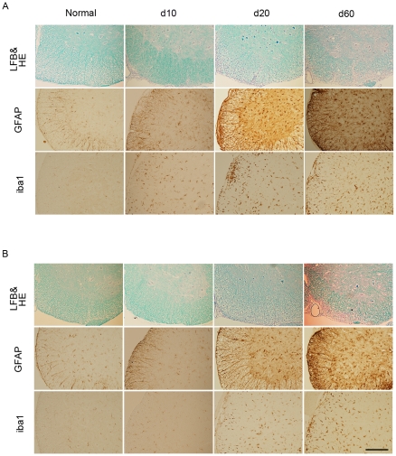
Figure 2. Histopathology of the spinal cords in EAE mice.
Representative histology of the spinal cords of WT (A) and Olig1−/− mice (B) in d10, d20 and d60. Lumbar spinal cords were stained with luxol fast blue (LFB) and hematoxylin and eosin (HE) (upper panels) and either an anti-GFAP (middle panels) or anti-iba1 antibody (lower panels). Scale bar: 200 µm and applies to all panels.

