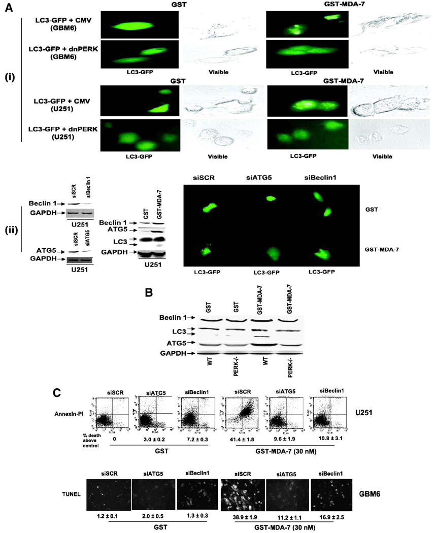
Fig. 2.
GST-MDA-7 causes LC3-GFP vesicularization in transformed cells in a PERK-dependent manner. A, (i), GBM6 and U251 cells were plated in four-well chamber glass slides in triplicate and 12 h after plating transfected with a plasmid to express LC3-GFP and in parallel co-transfected with either a vector control plasmid (CMV) or with a plasmid to express dnPERK. Twelve hours after transfection, cells were treated with GST or GST-MDA-7 (100 nmol/L). Twenty-four hours after GST-MDA-7 exposure, the GBM6 and U251 cells were examined under visual light (visible) or under fluorescent light (LC3-GFP). Representative images from the triplicate plating (n = 2). (ii), U251 cells were plated in four-well chamber glass slides in triplicate and 12 h after plating transfected with a plasmid to express LC3-GFP and in parallel co-transfected with either a vector control plasmid to express a nonspecific scrambled siRNA (siSCR) or plasmids to knockdown expression of Beclin-1 (siBeclin-1) or ATG5 (siATG5). Parallel studies also transfected cells with plasmids to express scrambled siRNA and untagged GFP. Twelve hours after transfection, cells were treated with GST or GST-MDA-7 (100 nmol/L). Twenty-four hours after GST-MDA-7 exposure, the U251 cells were examined under fluorescent light (LC3-GFP and GFP). Representative images from the triplicate plating (n = 3). Immunoblotting, cells transfected with siRNA constructs to modulate the expression of ATG5 and Beclin-1 were immunoblotted to determine the expression of Beclin-1 and ATG5 48 h after transfection. Cells treated with GST-MDA-7 and GST were immunoblotted 48 h after treatment to determine the expression of Beclin-1, ATG5, the cleavage status of LC3 and GAPDH (n = 2). B, Transformed MEFs (WT; deleted for PERK, PERK−/−) 24 h after plating were treated with GST or GST-MDA-7 (100 nmol/L). Twenty-four hours after GST-MDA-7 treatment, cells were isolated and subjected to SDS-PAGE to determine the expression of Beclin-1, ATG5, the cleavage status of LC3 and GAPDH (n = 2). C, GBM6 and U251 cells were plated in four-well chamber glass slides in triplicate and 12 h after plating transfected with either a vector control plasmid to express a nonspecific scrambled siRNA or plasmids to knockdown expression of Beclin-1 or of ATG5. Twelve hours after transfection, cells were treated with GST or GST-MDA-7 (30 nmol/L). Forty-eight hours after GST-MDA-7 exposure, the viability of the GBM6 and U251 cells was determined by: terminal deoxynucleotidyl transferase-mediated dUTP nick end labeling assay or; by Annexin V-PI flow cytometry on isolated cells (±SE; n = 3). Data shown are for GBM6 (terminal deoxynucleotidyl transferase-mediated dUTP nick end labeling) and U251 cells was determined by Annexin V/PI flow cytometry (±SE; n = 2). Data reproduced with permission from Yacoub et al. Caspase-, cathepsin and PERK-dependent regulation of MDA-7/IL-24-induced cell killing in primary human glioma cells. Mol Cancer Ther 7 (2008), 297–313.

