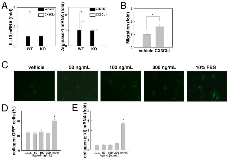
Fig 5. CX3CL1 induces alternative activation of Kupffer cells by expressing IL-10 and arginase-1 through CX3CR1.
Kupffer cells isolated from WT and CX3CR1-deficient mice were incubated with 100 ng/ml CX3CL1 or vehicle (PBS). (A) mRNA levels of IL-10 and arginase-1 were measured by quantitative real time PCR. (B) Serum-free media containing recombinant CX3CL1 (100 ng/mL) was placed in the lower chamber and Kupffer cells of WT mice were placed in the upper chamber. Migration of Kupffer cells into the lower chamber was counted 16 hours after stimulation. (C, D) HSCs of collagen promoter driven GFP transgenic mice were incubated with 50, 100 or 300 ng/ml recombinant CX3CL1 or vehicle (PBS) for 48 hours. (C) Representative photomicrographs of HSCs and (D) their quantification are shown (original magnification: ×200). (E) Collagen α1(I) mRNA are shown. Similar results were obtained in three independent experiments. *P<0.05.

