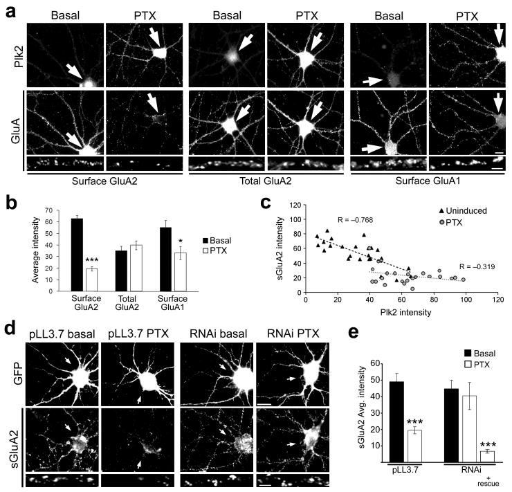
Figure 4. Plk2 induction is required for activity-dependent decreases in surface GluA2.
(a) Immunolabeling as indicated for endogenous Plk2 and surface or total GluAs in cultured hippocampal neurons under basal conditions and after stimulation with picrotoxin (PTX) for 24 h. Higher magnification views of representative dendrites are shown below. Arrows indicate cell body of neuron shown. (b) Quantification of data in (a). ***p<0.001, *p<0.05; N=25 neurons per condition. (c) Inverse correlation between Plk2 expression and surface GluA2 levels in individual cells under basal and PTX induced conditions. (d) Hippocampal neurons were transfected with pLL3.7 empty vector or Plk2 RNAi as indicated, along with pEGFP to mark transfected cells. Neurons were treated with PTX or vehicle (basal) and immunostained for GFP and sGluA2 as indicated. Arrows indicate transfected neuron cell body. (e) Quantification of data from (d). N=10–15 neurons per condition; ***p<0.001. Scale bars, 10 μm (wide view), 5 μm (magnified images).

