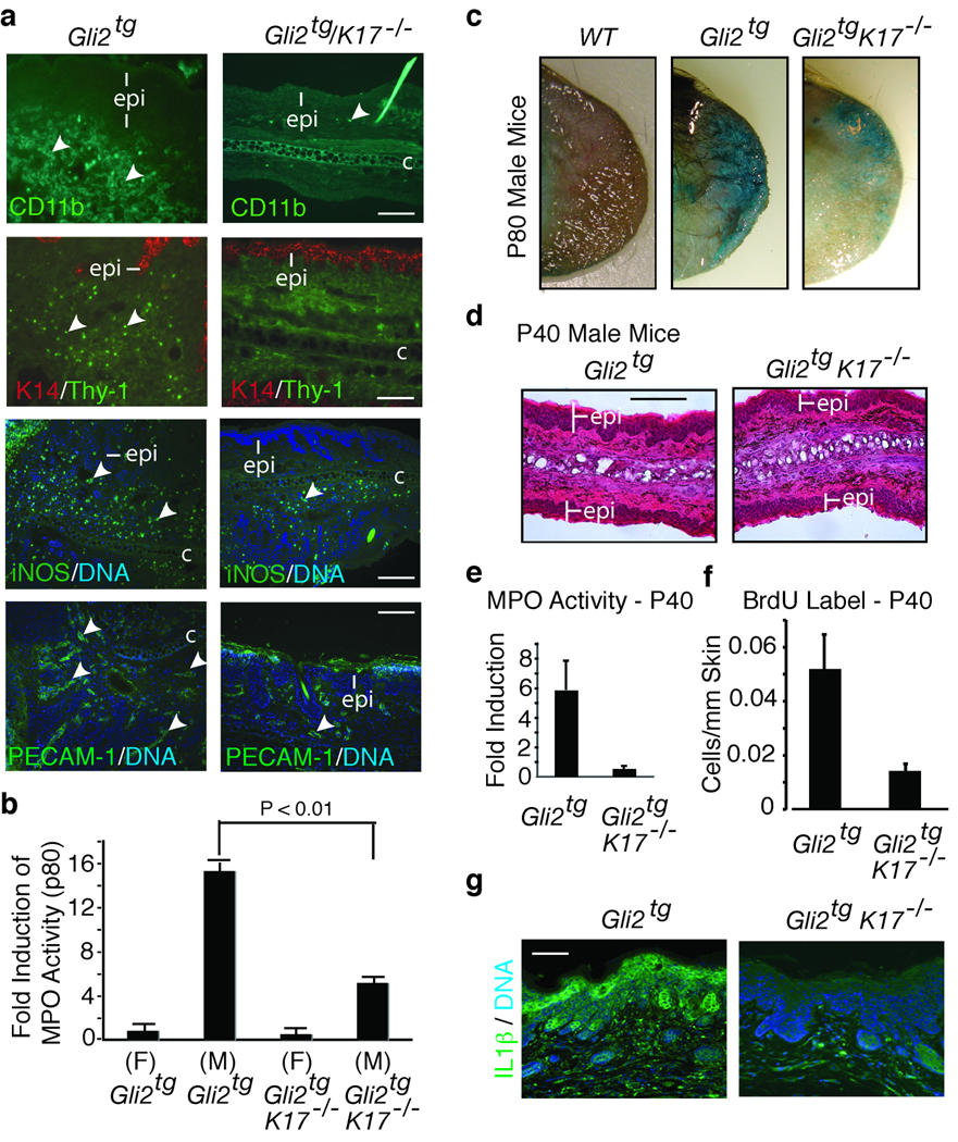
Figure 2.
Role of inflammation in the onset of ear lesions. (a) Immunodetection of infiltrating immune cells and vasculature in Gli2tg and Gli2tg/K17−/− male mice at P80 using antibodies to CD11b, Thy-1, iNOS, and PECAM-1 (see arrows). Labeling key provided in lower left corner. (b) Quantification of myeloperoxidase activity (MPO; mean ± s.e.m.) in ear tissue of mice at P80, normalized to female Gli2tg mice. (c) In situ beta-galactosidase staining in P80 male ear tissue of various genotypes. Blue staining reflects loss of barrier integrity. (d) Hematoxylin-eosin stained ear tissue of male mice at P40. (e) Myeloperoxidase activity in ear tissue of P40 male mice, normalized to female Gli2tg (mean ± s.e.m.). (f) Quantification of BrdU labeled cells/um of epidermis in P40 male mice. (g) Immunostaining for IL-1β in the epidermis (epi) of P80 male ear tissue. Scale bars: a (20µm), c (25µm), d (50µm).

