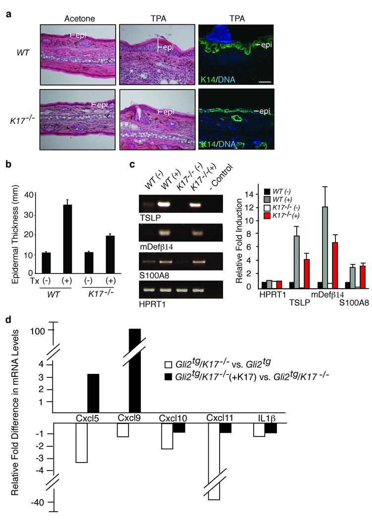
Figure 3.
Absence of K17 blunts epidermal hyperplasia and alters inflammation in a chemical model of dermatitis. Wildtype and K17−/− mouse ears were treated with acetone (vehicle control) or TPA. (a) Hematoxylin-eosin stained tissue sections depicting the effect of TPA treatment on thickness of epidermis. Right panel: Expansion of basal layer visualized by immunostaining for K14. Scale bars: 20 µm. (b) Epidermal thickness (mean ± s.e.m.), as conveyed by K14 staining, in vehicle (−) and TPA-treated (+) male mouse ears. (c) Right: Semi-quantitative RT-PCR survey of targets associated with loss of barrier integrity. Left: Quantitation of RT-PCR results shown in c. (d) Cytokine/chemokine expression in primary cultures of Gli2tg and Gli2tg/K17−/− keratinocytes 12 hours after TPA. For d and e, fold change represents changes due to loss of K17 (compilation of 3 assays involving distinct pools of mRNAs). (f) Changes in cytokine and chemokine expression in Gli2tg/K17−/− after reintroduction of K17 via transfection (triplicate).

