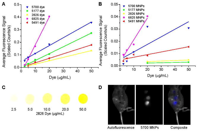
Figure 2.
Fluorescence of NIR dyes in vitro (A) and dye-loaded MNPs in vivo (B). A small amount (3 μl) of each hydrophobic dye in ethanol was dropped onto filter paper and the fluorescence intensity measured with manually drawn ROIs. Magnetic nanoparticles containing 0.25–5.0% w/w dye were suspended in mannitol citrate buffer (1 mg MNPs/mL) and 20 μL was subcutaneously injected and immediately imaged. Representative images are shown for dye 2826 in ethanol (C), and a mouse subcutaneously injected with MNPs loaded with dye 5700 at 0.25, 0.5, and 1.0% w/w dye in 1 mg/mL MNPs (D).

