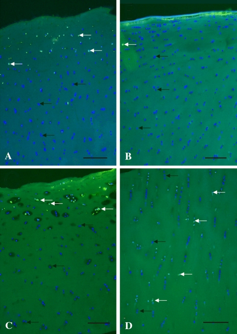
Fig. 5A–D.
An histologic assessment of the distribution of chondrocytes in articular cartilage explants was performed. The TUNEL assay (FITC-Annexin-V) with DAPI counterstaining shows apoptotic cells after 24 hours of treatment in osteochondral ex vivo specimens. The results of (A) lidocaine, (B) betamethasone, (C) prednisolone, and (D) prednisolone-lidocaine combinations are shown. The blue staining shows all nuclei of living cells (black arrows), illuminating green (TUNEL-positive) cells are dead (white arrows) (Original magnification, ×50). Scale bar = 150 μm.

