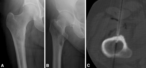
Fig. 1A–C.
(A) AP and (B) lateral radiographs are shown of the right hip of a 52-year-old woman with progressive, localized right thigh pain occurring at rest and at night . She has no history of cancer but is a long-time cigarette smoker, and a CT scan of the chest revealed a lung nodule and a thyroid mass. Although the plain radiographic appearance is suggestive of fibrous dysplasia, an alternative cause for her pain was not identified, therefore, a CT-guided needle biopsy was performed. (C) The biopsy revealed “bone and fibrous tissue with reactive changes” per the formal pathology report. There was “no malignancy in the specimen”. In this case a definitive diagnosis of fibrous dysplasia was not provided by the pathologist, but the surgeon chose to counsel the patient and continue to follow her with serial radiographs. In the biopsy database, this lesion was categorized as fibrous dysplasia.

