
Figure 1B.
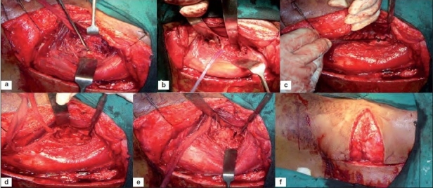
Intraoperative photograph of exposure shows (a) blunt dissection done in front of transverse process creating an anterior flap of muscle of psoas, quadratus lumborum (b) lumbar nerves identified, protected (c) A spatula was placed under reflected psoas muscle exposing anterolateral surface of fractured vertebral body (d) spinal cord decompression done by corpectomy of fractured vertebra and removal of adjacent disc and bed for graft created (e) tri-cortical strut graft from ipsilateral iliac crest, placed between fractured vertebral body and proximal intact vertebral body (f) wound closed in layers