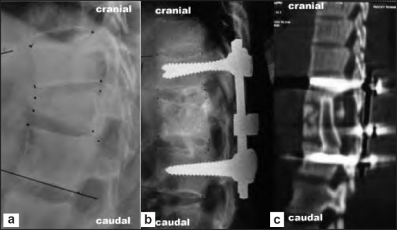
Figure 3.

(a) Preoperative X-ray of dorsolumbar spine lateral view of 32 years old male with burst fracture (type A) of D12 with preoperative kyphosis of 32°. (b) Immediate postoperative X-ray lateral view after single-stage anterior decompression, bone grating, and pedicle screw fixation via extrapleural retroperitoneal approach, showing correction of kyphosis and well-placed bone graft. (c) Six months postoperative migsaggital reconstructed CT image showing graft incorporation and final kyphosis of 11°
