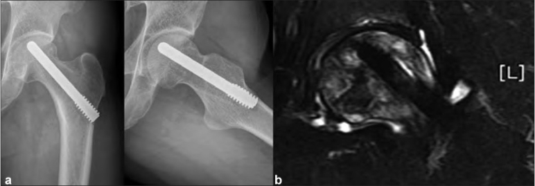
Figure 1.

(a) Postoperative radiograph anteroposterior and lateral views showing a well placed implant with no signs of subchondral collapse or depression in the articular surface. (b) Follow-up MRI scan showing porous tantalum rod in the necrotic area, with reactive marrow signal changes around the tip of the implant, without any evidence of femoral head collapse
